Saturday, August 17, 2024

BLUE Protocol

BLUE Protocol was developed by Lichtenstein et al (Chest 2008) as an tool to asses the cause of respiratory failure using ultrasound .

Probe placement


This method defines three standardised points: (i) The upper BLUE-point is at the middle of the upper hand; (ii) The lower-BLUE-point is at the middle of the lower hand and the (iii) The PLAPS-point (posterolateral alveolar or pleural syndromes), which is indicated by the intersection of a horizontal line at the level of the lower BLUE-point and a vertical line at the posterior axillary line.

  • They had developed an algorithm - Bedside  Lung Ultrasound in Emergency and was studied retrospectively with an accurate diagnosis of 90.5% of the cases. 


Updated on 17/8/2024
Reference
  • Lichtenstein DA, Mezière GA. Relevance of lung ultrasound in the diagnosis of acute respiratory failure: the BLUE protocol. Chest. 2008 Jul;134(1):117-25. doi: 10.1378/chest.07-2800. Epub 2008 Apr 10. Erratum in: Chest. 2013 Aug;144(2):721. PMID: 18403664; PMCID: PMC3734893.

What is Curtain sign, quad sign ?

Curtain Sign

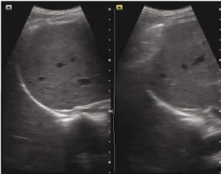
  • Curtain sign is a sonographic characteristics of the inferior most part of the lateral lung which occupies the costophrenic recess. 
  • Curtain sign in normal lung will demonstrate 
    1. Dynamic movement of the lung curtain which is due to expansion and retraction of the lung (Lung curtain moves up and down).  
    2. Lateral aspect of the diaphragm and the upper abdomen is always covered/obscured by the lung curtain, irrespective of the phase of the respiratory cycle.

  • Absence of either or of the above results in curtain sign which is commonly seen in pleural effusion.

Curtain sign – the aerated lung in the costophrenic recess overlaps part of liver and diaphragm creating a demarcated leading edge of the lung air artefact, giving the appearance of curtain (of air). Both images are in different part of the respiratory cycle

Click here for the video : Curtain Sign / Abnormal Curtain Sign
    

Quad sign

In case of pleural effusion, the pleural layers are separated by fluid in between, the visceral pleura appears as a line (the lung line), which is regular and almost parallel to the parietal pleura (pleural line). Together with shadows from the adjacent rins, these lines draw a four sided figure which is known as the Quad sign .

Sinusoid sign

When examined on the M-Mode, a sinusoid pattern is seen which is due to the movement of the lung line towards the pleural line on inspiration and back on expiration. This is called as a sinusoid sign. 


A. quad sign: The area of pleural effusion is framed within four regular borders: the pleural line-P, the shadow of the ribs (stars), and the almost regular deep border, which is the lung line-L; B Sinusoid sign : M-mode image of pleural effusion.
   

Friday, August 16, 2024

What is TAPSE ?

  • Tricuspid annular plane systolic excursion (TAPSE) is a simple and reproducible parameter for quantitative assessment of the RV EF. It was first described by Kaul at al (Am Heart J. 1984).
  • It is perfomed from an apical four chamber window using M mode across the annulus to evaluate longitudinal contraction.
    • TAPSE >16mm is considered normal.
    • TAPSE< 16mm indicates reduced RV Ejection fraction. 
  • AHA currently recommends TAPSE as one of the tool to assess RV Function. It is an independent prognostic marker in PE, IWMI, ARDS. 
  • Alternative to A4Cview subcostal view may be used in mechanically ventilated patients. 


Reference

  • Kaul S, Tei C, Hopkins JM, et al. Assessment of right ventricular function using two-dimensional echocardiography. Am Heart J 1984;107:526-31. 10.1016/0002-8703(84)90095-4 
  • Main, A.B., Braham, R., Campbell, D. et al. Subcostal TAPSE: a retrospective analysis of a novel right ventricle function assessment method from the subcostal position in patients with sepsis. Ultrasound J 11, 19 (2019). https://doi.org/10.1186/s13089-019-0134-7